Bottom jaw implantation case
This is a 68-year-old male patient.
I came to the hospital because it was discovered in the lower anterior teeth.
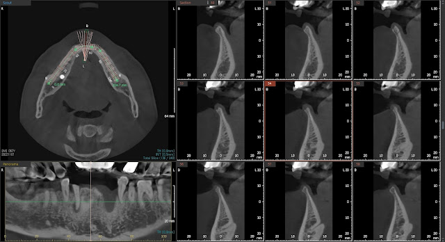
This is a CT finding.Bone defects are visible in the area where implantation is intended.
This is a photo while drinking.
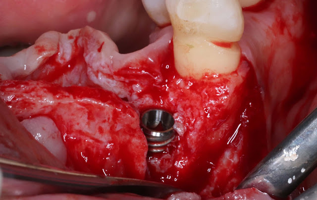
SBA 4.0 was installed.Dehiscence defect is visible.
We performed bone grafting using a xenograft mix and Xenoguide, an absorbable membrane.
This is a postoperative radiograph. Bone healing 503 was inserted and GBR was performed.
3 months after surgery. This is a photo and a photo after the final prosthesis.Because the initial fixation was not very good, immediate loading was not performed.
The final prosthetics was done 4 months ago.


Comments
Post a Comment