Posts

Showing posts from July, 2022

The removal method for implant fracture.

Image
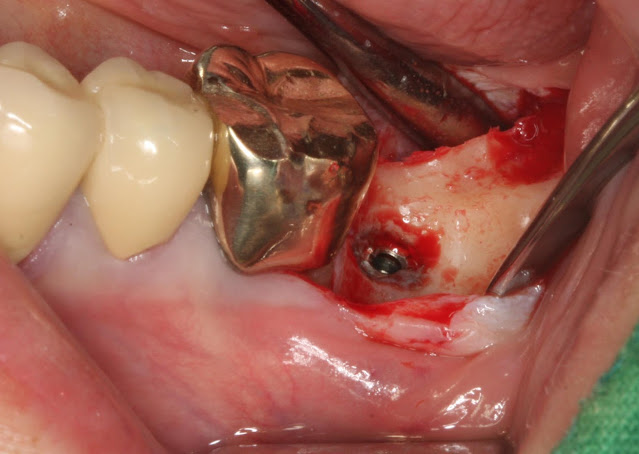
The usual method for removing a fractured implant is as follows. The implant has fractured. In the case of the mandible, it seems that there are many instances where the implant cannot be easily removed even with a fixture removal kit. In such cases, a trephine bur with a slightly larger diameter than the implant is often used to drill at least half of the length of the implant. Of course, it is essential to proceed with caution to avoid any nerve damage during the procedure. Afterward, using a root picker or a thin elevator with gentle force in a mesiodistal direction, the fractured implant usually starts to move slightly. If the implant does not move at all, then re-drilling deeper with the trephine bur and attempting again usually results in successful removal. After the removal, the implant was re-implanted. (This is not a healing abutment; it is a picture of the abutment taken during prosthetic work.) It seems that both of us frequently encounter implant fractures or tearing, both

Implant placement through tooth extraction socket.

Image
The patient is a 79-year-old male who came for dental treatment due to fractured teeth in the maxilla at tooth numbers 13 and 15. The radiograph provided is an image taken before the fracture occurred. Based on the CT findings before the fracture, tooth number 13 showed a position toward the contralateral side. Tooth number 13 was extracted by carefully placing the drill on the palatal side of the root and directly drilling to remove the tooth cautiously. Implant placement for tooth number 3 was done on the palatal side because the contralateral alveolar bone was thin, making it a suitable location for the implant. Bone grafting with allograft was performed on the contralateral side during the extraction. After maintaining the resorbable membrane, the suturing was performed to minimize the exposure of the contralateral flap while keeping it protected. One week later, the sutured area has opened up. It could be due to suturing too tightly or may be influenced by factors such as smoking.

Upper jaw bilateral implants.

Image
The patient is a 58-year-old male. The implant placement is scheduled to be done on both sides of the maxilla (upper jaw). On the left posterior region, a crestal approach implant was placed. And on the right first premolar, which had severe periodontitis, we extracted it first. After about 4 months of extraction, we are planning to place an implant in the right premolar area. Also, there is a defect in the alveolar bone in the area of tooth number 14. In the area of tooth number 14, we first performed a simple bone healing using Bone Healing 503 to prepare for Guided Bone Regeneration (GBR). After the allograft (bone graft from a different individual) transplantation, we covered it with a resorbable membrane. The healing abutment was placed to serve as a support structure and facilitate the process. It has been sutured.   This is the post-surgery radiograph. Q. After using the resorbable membrane, how did you fix it in place? I haven't had good results, so recently, I mainly use T