Posts

Showing posts from November, 2022

A case of maxillary sinusitis due to peri-implantitis

Image
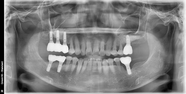
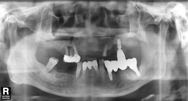
This is a case of maxillary sinusitis caused by periimplantitis in a 64-year-old female patient this year. The patient had undergone a sinus membrane elevation procedure for implant placement in the maxillary molar area about 20 years ago. Here is an intraoral photo from 2004. Here is a photo of the same patient undergoing a window approach in 2004. First, bone grafting was performed using Bio-Oss. Implants were placed approximately 6 months later, and this is a panoramic image from 2007 after the prosthetic restoration. It's 2009 It's 2012 It's 2014 It's 2017 It seems there were no significant issues at this time in 2020 as well. Here is the picture from 2022. Suddenly, not only around the implant in the 17th area but also in the 36th area, periimplantitis has developed, leading the patient to visit. The area around implant number 17 appears to have developed periimplantitis, with a deep pocket extending almost to the maxillary sinus floor.   Compared to the area aro

Tooth extraction due to periodontal disease

Image
This year's patient is a 67-year-old woman. The maxillary left first molar was extracted due to periodontal disease. A photo taken approximately 3 months after the extraction. Despite the limited remaining bone, I have decided to proceed with a crestal approach for implant placement. Here is a photo immediately after the surgery. Here is a photo after approximately 3 months post-surgery. Here is a photo taken shortly after the restoration approximately 5 months post-surgery.  

Immediate implantation case

Image
Shall I share a brief case of immediate implant placement today? A 79-year-old male patient with chronic periodontitis in the #47 region, exhibiting significant mobility but not severe bone resorption, underwent extraction with immediate implant placement due to the mobility issue. This is the CT finding. I placed the implant with some concern while extracting the tooth. For immediate implant placement in the mandibular second molar area, it seems advantageous to place the implant slightly toward the distal aspect with some caution, considering the future restoration's emergency profile. The implant is completely submerged within the bone, and it was placed with an approximate depth of around 4mm to achieve a proper gingival height. Collagen plug was used to prevent the penetration of foreign substances without performing separate bone grafting.  Afterward, no sutures were applied, and Periopack was maintained. Bone healing abutment (size 604) was placed to avoid interference with

Implant placement in a patient with osteoporosis

Image
A 59-year-old female patient with a history of osteoporosis medication for about 3 years and a drug holiday of approximately 6 months for implant placement. Based on the CT findings, the bone quality of the lower left posterior area appears to be similar to D4 and the bone width also appears narrow. During the surgery, a radiograph was taken showing that the initial stabilization of the implant in the area of tooth #36 was not successful, causing it to continuously shift downward into the bone. This has made the removal of the implant quite challenging, and there is a risk that it could further embed itself deeper into the bone if not managed carefully. With no other option, the cortical bone on the buccal side was cautiously removed using a round bar, and the implant was delicately extracted using forceps.   Subsequently, allograft was transplanted into the bone, and the implant was re-implanted cautiously. Guided bone regeneration (GBR) was also performed on the buccal side. Such sym

Peri-implantitis due to diabetes and poor condition

Image
This is a photo from the initial examination in 2013, with a 72-year-old male patient. Four implants were placed in the maxilla, and a bar over denture was fabricated as shown in the picture. The existing implants were left in place for now, as the patient did not wish to remove them. Intraoral photo. This is a photo from 2016. This is a photo from 2018. At this time, the existing left implants were removed. In the 2020 photo, signs of peri-implantitis are visible around the implants on the right side. The patient appeared to have progressed diabetes and poor oral hygiene. Bone grafting was performed after thorough cleansing using a titanium brush and detoxification with TC (tetracycline). After suturing: Preoperative radiograph: Postoperative radiograph: One-year postoperative photo: The patient's discomfort has decreased, but complete bone formation as expected has not yet occurred. However, this time there is pus coming out on the left side, and the gingiva is showing signs of s