Posts

Showing posts from June, 2022

Immediate implant placement case 1

Image
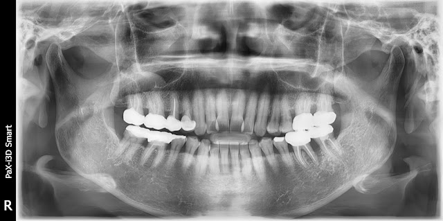
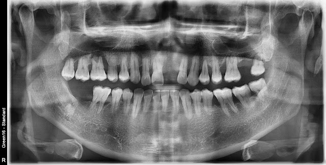
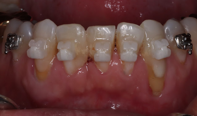
The patient is a 46-year-old male. The patient is experiencing persistent discomfort in the area of tooth number 12. After the extraction, implant placement is being planned. The tooth mobility was severe, so the decision was made to extract it and immediately place an implant during the same procedure. Tooth number 11 was extracted and an implant was placed simultaneously while performing root canal cleansing. An implant was placed, and at the same time, contouring augmentation was performed using allograft during the procedure. This is the picture after suturing. After placing a healing abutment, bone grafting was performed around it, and the area was sutured in a coronal direction. This is a picture taken approximately 4 months later with a temporary prosthesis attached. This is a picture with the final prosthesis attached. The picture after one year has passed.  

Damaged distal root.

Image
This surgery was performed in the year 2015. The patient is a woman in her 60s who came for a dental visit due to discomfort with tooth number 46. Tooth number 36 shows unfavorable conditions in the distal root area. Tooth number 36 shows unfavorable conditions in the distal root area. Tooth numbers 36 and 37 are connected by a prosthesis, and there is no mobility. After discussing with the patient, it has been decided to remove only the distal root for now. During the surgery, the level of root resection was determined and performed while monitoring the cutting of the root apex. The root resection was performed to ensure effective cleansing, allowing for thorough cleaning in the end. This is the post-procedure radiograph. This is the radiograph taken approximately 7 years after the procedure. There are no specific issues, and it is functioning well.

When there is damage to the alveolar bone

Image
The patient is a 44-year-old male. He is considering implant placement in area 16, but there is significant damage to the alveolar bone. In this case, socket preservation was performed simultaneously with the extraction. The pre-extraction condition. I have confirmed the pre-extraction condition. Symptoms of periapical infection are evident, and the maxillary sinus membrane appears to be thickened. The extraction was performed simultaneously with bone grafting using allograft and collagen. The pre-implantation condition approximately 5 months later. The pre-operative condition is shown here. The thickening of the maxillary sinus membrane appears to have disappeared. Based on this level of healing, it seems that a crestal approach implant placement may be feasible. The post-operative condition after crestal approach implant placement and bone grafting. In approximately 2 months post-operation, here is the picture. This is a radiograph taken during scanning Here is the picture taken duri

Bone Healing Abutment Implantation Case

Image
Here's a case of a 70-year-old male patient  Implantation was immediately planted in area 46. Normal healing abutment was installed after SB bone level  fixture  was implantated in the usual way. However, the radiation shot indicates that the healing abutment is not completely locked. In this case, if you lock it with OSSTEO BIONICS' Bone Healing Abutment, you can easily see it locked without interference from the adjacent bone. I think this type of healing is very convenient for immediate implantation or GBR. It makes soft tissue more comfortable and fits well without interference from neighboring bones. It's a month before the final prosthesis. We change the abutment to the type of healing abutment that is needed. This is the final prosthesis shot. Bone Healing Abutment seems to be advantageous for implantation for people with soft tissue like this, even during simultaneous GBR for mandibular cavity.

Extraction and extraction with preservation method.

Image
  This is a 43-year-old male patient. I'm planning to implant my tooth number 27 because it's not in good condi tion. The degree of agitation of the teeth is severe and the loss of the narrow alveolar bone is observed.  In this case, it seems that there are many cases where the implant is implanted with extraction and preservation at the same time as removal. After extraction, bone transplantation was performed and covered using collagen. It's about 4 months later. About 5 months later, the implant was planted. I think the alveolar bone is enough to implant. This is a suture after surgery. Additionally, contourig augmentation was implemented. Radiography after extraction and preservation. This is the final picture after prosthesis.