Posts

Showing posts from September, 2022

Implant placement in a patient with chronic periodontitis

Image
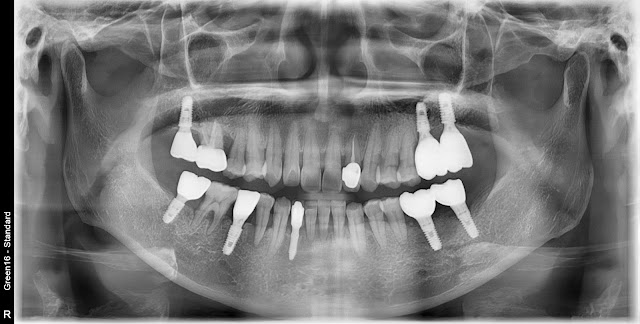
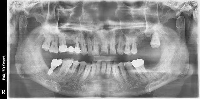
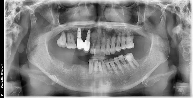
A 59-year-old female patient, immediate implant placement is planned for the site of tooth #46. The tooth was extracted due to severe tooth mobility caused by chronic periodontitis. There is partial loss of alveolar bone on the adjacent side, but it doesn't appear to be severe. An implant was placed, and Collacera was grafted in the gap area. A bone healing abutment was attached. Periopack was placed in the upper area. This is a postoperative radiograph. This is the final restoration photo approximately 4 months after the surgery.  

Maxillary sinus infection

Image
In this post, I will upload the treatment process for the infected case after the Windows app. This is a male patient in his mid-50s. A window approach was performed in the left maxillary sinus, but the patient was referred due to symptoms of infection, including the presence of pus in the surgical site. Based on the CT findings, signs of infection are evident in the left maxillary sinus. There is a history of medication for over 3 weeks, but there was no improvement. Irrigation of the left maxillary sinus was performed through the oral cavity by inserting a drain as shown in the image. During this procedure, a significant amount of pus drainage was observed. Irrigation of the maxillary sinus was performed twice a week with a one-week interval, and it was confirmed that no pus was coming out. The drain was subsequently removed. During irrigation, some of the transplanted bone came out, but the internal bone was intentionally not removed. A month later, in the photo, there are no sympto

Socket Presevation

Image
This year, a 63-year-old male patient presented with severe periodontitis in the mandibular left posterior region. Socket preservation was performed immediately after tooth extraction using a non-resorbable membrane and allograft bone material. Oral intraoral photograph. This is the incision and reflection of the tissue to access the underlying tissue after tooth extraction for the purpose of utilizing the surrounding tissue. After thoroughly removing the infected tissue in the lower jaw, homogenous bone graft was performed, followed by placement of a non-absorbable membrane and suturing. A non-absorbable membrane, specifically an open-textured membrane, was used. About 2 weeks later. About 4 weeks later. Removed the membrane Here is the appearance after approximately 4 months. This is the picture before implant placement at approximately 5 months. Additional guided bone regeneration (GBR) was performed using an absorbable membrane due to the presence of slightly soft and insufficien

Concerns about nerve damage and narrow bone width during implant placement

Image
Last week, dental implants were placed in the lower right molar area of a 61-year-old female patient. Due to the narrow bone width, especially in the premolar area, I recommended using a bone-level implant such as the Roxolid implant. However, due to cost constraints, we decided to proceed with a tissue-level implant instead. Taking into consideration the limited mouth opening of the patient, as well as the occlusion and the relationship with the inferior alveolar nerve, a mid-face X-ray was captured. Subsequently, implants with a diameter of 3.5mm were placed in the area of tooth #45, while the rest were 4.0mm in diameter. The implants had a tissue-level neck of 2.8mm and a length of 8.5mm. The implant placement was accompanied by guided bone regeneration (GBR) techniques. However, there is a concern for potential nerve damage in the area of tooth #47, contrary to initial expectations. Here is a post-operative photo. Fortunately, the area of tooth #47 was placed slightly towards the l