Posts

Showing posts from October, 2022

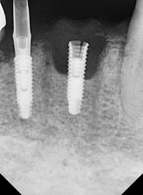
The mobility of tooth #11 and bone loss

Image
A 56-year-old male patient. The patient presented with pain and discomfort in the area of tooth #11. The alveolar bone on the palatal side shows signs of resorption, but the condition appears relatively stable. Adequate socket debridement was performed after tooth extraction. Collagen material was briefly grafted into the gap area. Preoperative radiograph before the surgery. Postoperative radiograph after the surgery. Bone healing abutment has been placed. Here is a photo taken about 2 months later. The thick gingival biotype suggests a favorable prognosis in this case. After the final restoration  

Re-surgery following implant fracture

Image
This year, a 74-year-old male patient visited us with a mini dental implant that had been placed in the lower jaw. After 4 years of prosthetic use, one of the implants came loose and torn as seen in the picture. The patient had a bar overdenture in place over the edentulous ridge. These days, mini implants and similar approaches are receiving attention in the trend of minimalism. In this context, one of the primary considerations should be to provide sufficient strength. Before the surgery Implanted simultaneously with GBR four years ago A week after the surgery. 2 weeks after. 4 months after After the completion of the final prosthesis. But, after 4 years, eventually.  W e need to start over from the beginning.

Simultaneous mandibular resection and bone grafting (BGR).

Image
The patient is a 55-year-old female with ameloblastoma in the left mandibular angle area. Mandibular resection was performed along with simultaneous iliac bone grafting. Here is the current state of implant placement after the procedure. the photo is not of the best quality due to its age. This is a photo from 2004. This is from 2008 This is from 2011 This is from 2021 It's 2021 too.   These days, although not commonly used, it seems that external hex type implants have advantages when creating multiple screw-type prosthetics.

A case with a less favorable condition of the palatal root.

Image
This patient is a 51-year-old male who presented with pain in the area of tooth #26. Based on the CT findings, due to the unfavorable condition of the palatal root, we performed socket preservation using allograft bone and a collagen plug during the extraction of tooth #26. After the extraction. Subsequent to approximately 4 months of waiting, here is the photo. It appears that the palatal root area has shown some degree of bone formation. I placed an Ossteo Bionics SB implant flaplessly. 4 months after implant placement.   Proper socket management in the maxillary posterior region can potentially simplify the surgical procedure.