Posts

Showing posts from December, 2022

A case of guided bone regeneration (GBR) in the left posterior region.

Image
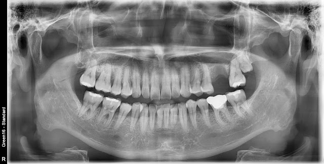
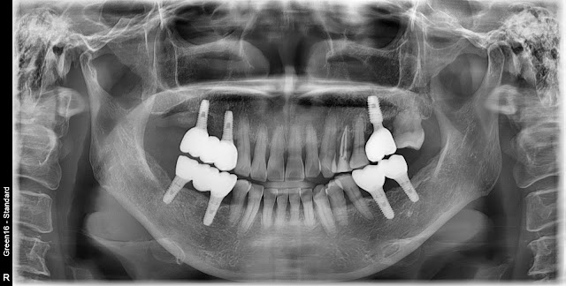
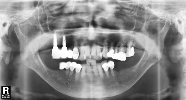
This is a case of a 75-year-old female patient who underwent a relatively simple GBR procedure in the mandibular left posterior area due to factors such as the patient's age and fear of surgery. The height of the alveolar bone in the mandibular left posterior area is sufficient, but the bone width appears to be thin. In such cases, there are various methods that can be considered. However, I first performed a simple guided bone regeneration (GBR) procedure using resorbable membrane and bone grafting material. I exposed the connective tissue, made a vertical incision, and performed homogenous bone grafting. Afterward, I covered the area with a cross-linked resorbable membrane. During the vertical incision, it's important to be cautious and avoid any damage to the mental nerve. This is a photo taken about 4 months after the surgery. Post-operative radiograph after the surgery. A combination of allograft and autograft was used for bone grafting. Implants were placed approximately

Case of natural tooth extraction and implant placement

Image
The patient is a 48-year-old male who visited after the natural extraction of tooth 26. According to the CT findings approximately 3 months later, mucosal thickening is observed in the left maxillary sinus, and the remaining alveolar bone seems suitable for a crestal approach. With the localized mucosal thickening observed in the left maxillary sinus, predominantly in the lower region, I have decided to proceed with crestal bone grafting and implant placement. After surgery This is the result about 3 months after the surgery. This is the outcome approximately 4 months after the initial surgery and the subsequent secondary procedure. After final prothetics. When performing a sinus lift procedure, it's important to evaluate whether there are any pathologic changes within the maxillary sinus and if possible, assess whether drainage through the middle meatus is functioning properly on CT imaging.  

Case of chronic periodontitis.

Image
A case of immediate implant placement in a 59-year-old female patient with chronic periodontitis involving teeth #24 and #25. There is some alveolar bone loss visible. The tooth was extracted and an immediate implant placement was performed. The initial stability was satisfactory. After about 4 months, the prosthesis was placed.

Case of implant placement in a narrow ridge

Image
A 74-year-old female patient this year. This is a photo from 2013. Planning to place implants in the lower left posterior area. The width of the bone ridge appears to be too narrow for procedures like ridge splinting. Consequently, a decision was made to perform Guided Bone Regeneration (GBR) first, followed by a secondary implant placement. GBR was initially carried out using allograft bone material through the use of SmartBuilder technology. The current appearance approximately 5 months after the surgery is as follows. The implant has been placed. Although the bone's shape hasn't completely remodeled, there were no significant issues with the implant placement. A secondary surgery was performed approximately 4 months later. The final prosthetic photo. A photo taken about 9 years later. CT findings approximately 9 years later. Immediate implant placement was performed using bone healing in the upper left molar region of the same patient. This is the appearance after placing th