Posts

Showing posts from January, 2024

Case of implantation of person with diabetes

Image
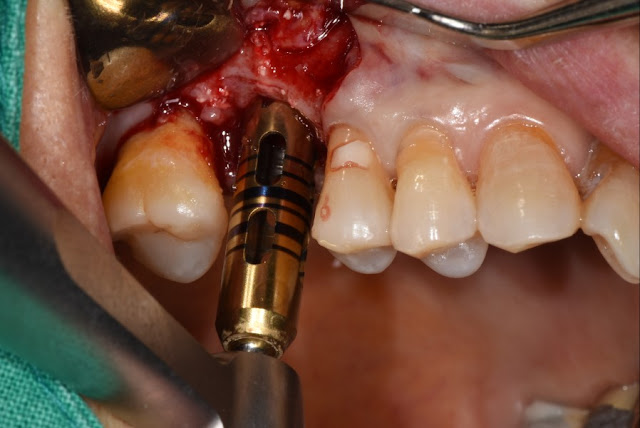
This is a 74-year-old male patient.  MS implants were placed in the lower anterior teeth. Here is a patient with poorly controlled diabetes. This is a photo before and after surgery. This photo is one week after surgery and the photo is about one month after surgery. The implant on side #41 showed signs of infection, was painful, and had some mobility, so it was removed. This is the picture one month after removal a nd this photo is about 5 months later. The area where the implant was removed appears to be healing. The implant is being placed again about 5 months after extraction. This photo is taken 4 months after implantation. This is a photo after the final prosthesis.

Multiple fixture installing case

Image
I removed the tooth using a trephine bur. A few months later, it will be replanted with a slightly larger fixture. When installing multiple fixtures, use a guide in advance to estimate the expected position of the fixtures before incision. This is the last detention.  We selected the location using a guide.  It's convenient even if you can't see.  

Apex tooth problem case

Image
There is a problem with the apex of the tooth. I remove the damaged tooth carefully. After waiting for the soft tissue to heal, the main level fixture is placed. There is no special reason. It was chosen considering the distance between natural teeth. No materials or additional procedures that could become foreign substances inside the extraction site were used, and only soft tissue suturing was performed.

Case of ossteointegration loss

Image
This is a photo of an implant that was used for over 10 years and was removed due to loss of osseointegration.This is a photo from 2011. Tissue level implants are functioning in areas #25 and# 26 of the lower jaw. These are photos from 2014 and 2018. This is a 65-year-old female patient.These are photos from 2021 and 2022. If you look closely, an abnormal reaction is observed around implant number 26, but the patient did not have any specific symptoms. The occlusion is mandibular #25 #26 is slightly under occluded, so only the prosthesis will be manufactured again in the future. However, patient came to the hospital today with a sore feeling when biting. Radiological findings show signs of loss of osseointegration in area 26. In the end, the implant was removed and a 5.0 tissue level implant was placed.We don't know what the exact cause was, but if it fell, patients looses of osseointegration like this.